Wednesday 21 April 2021

A new method of surgically treated nerve lesion recovery is used in Iceland

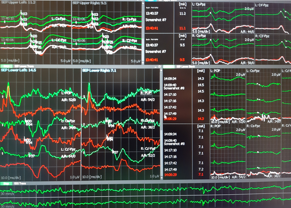
Peripheral nerve injury recovery is assessed in small mammals with nerve conduction studies 

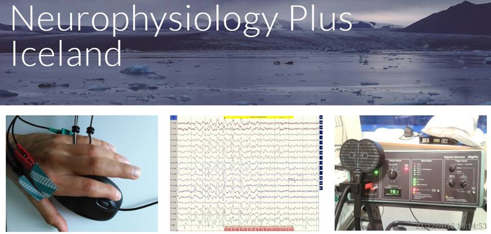
Possible research work with an Icelandic company which promotes technologies of regenerative biological tissues, the use of fish skin and fatty acids in the cellular-therapy

Ovidiu C. Banea
21.04.2021, Reykjavik

Motor neurography of the sciatic nerve to right gastrocnemian muscle was used successfuly in a laboratory mouse (Mus musculus) and a laboratory white rat (Rattus norvegicus) on 21st of April 2021. The technique was performed by Ovidiu C. Banea (neurophysiologist) and the Aron Jónasson (biologist) at invitation of Svava Kristinsdóttir, a research scientist from Kerecis and Dr Ingvar H. Ólafsson, neurosurgeon who collaborate with the company.

The nerve microsurgery (microscope) and preparations were performed by Dr Kristín Lilja Eyglóardóttir (Neurosurgeon) and Dr Baldur Tumi Baldursson (Dermatologist). 

The "in vivo" motor neurography (nerve conduction study) in an anesthetized laboratory small mammal, might be a summation as a quantitative recovery measurement in a broader Icelandic research project. 

Motor neurography, Mus musculus 10-20 minutes after e.

CMAP from gastrocnemian muscle of euthanized Mus musculus,
Amplitude 35uV, Latency 2.5 ms.

Performing technique in euthanized Rattus norvegigus

Dr Kristín Lilja Eyglóardóttir, 
performing end-to-end neurorrhaphy of sciatic nerve "in vivo", 
Rattus norvegicus, anesthesia with Isoflurane

It seems that end-to-side suture and end-to-end suturing appear to result in equivalent axon regeneration at 12 weeks, end-to-end at 6 weeks showing better axon regeneration rate, which may reduce target organ muscle atrophy (Tateshita et al, 2018).

Friday 13 December 2019

Road map for TMS and schizophrenia project in Iceland

Reykjavík University, Iceland

by Ovidiu C. Banea

On June 2018 a project dedicated to schizophrenia patients with auditory verbal hallucinations and their treatment with trans-cranial magnetic stimulation started in Iceland. 
The perfect coordination between researchers from Kleppur Psychiatric Hospital, Biomedical and Neural Engineering Institute at Reykjavík University, School of Business - Department of Psychology, National Institute of Neurological Disorders and Stroke (NINDS) Bethesda, United States and Clinical Neurophysiology Unit from National University Hospital - Landspítali Iceland made possible the recruitment and treatment of 12 patients with consequent analyses with multi-metric approach.
Measurements of the brain activity was performed before and after the treatment with P50, N100-P300 complex, resting state and auditory motor-task with 256 High Density-EEG recordings, cortical silent period and psychometric scales.
The team could organize and present their methodology, results and preliminary data in 14 original works during several local and international meetings in France, Japan, Poland, Portugal and Austria (see all works here).


On 13th of December we met at Reykjavik University and we discussed the detailed of the most important aspects of the AVH-TMS Icelandic Clinical trial - publishing the results in a peer review international journals. Two teams were formed, one at Reykjavik University and one at University National Hospital. The first team will process event related potential results at BNE and the second team will put together all data obtained at Fossvogur Landspítali and at Kleppur Psychiatric Hospital. By the end of January 2020 the process structure and software analyses will start.

Friday 1 November 2019

Assessment of the lower limb weakness and suspected amyotrophic lateral sclerosis

A patient with suspected motor neuron disease (MND or ALS) was assessed with TMS in our Unit in Iceland here. There were no signs of EMG criteria. We further investigated with TMS and it seems that there is a lower spinal level delay of the central motor conduction.


Monday 8 April 2019

FIRST HEILA VISTFRÆÐI JOURNAL CLUB MEETING

Possible evidence of human ability to detect Earth's magnetic field found HERE.
First Journal Club Meeting at Reykjavik, Iceland

We talk about the 6th human sense, MAGNETO-RECEPTION IN HUMANS


Thursday 24 January 2019

D wave IONM in Iceland. Third case: Meningocele


IONM for medular herniation at T5 level Use of D-wave 

HERE

Ovidiu C. Banea, Halldór Skúlasson, Ingvar H. Ólafsson, Aron D. Jónasson and Eysteinn Ívarsson

52 y.o. with meningocele.

Modalities:

MEP with direct cortical stimulation (560 V to the left and 890 V to the right) with train of five from C1-C2 and C3-C4 to:
- Right EDC, APB, TA, AH
- Left APB, TA, AH
SSEP were performed from lower tibialis nerve and recorded to FpZ-Cz´ and from median nerves to Fpz-C3´ and Fpz-C4´. Both were controlled at popliteal fossa level (TN) and spinal C7 (TN and MN).
D wave was obtained rotral and caudal to the defect with D-wave electrodes after stimulation at 1Hz continuously (199V)
TOF was used from rioght median nerve to right APB.  was 100-99% during the entire surgery.
EEG was analized from C4´-Fpz and Fpz-C3´channels.

Results:
At the beginning of the surgery MEP was obtained in the upper limbs and left lower limb muscles. Right AH muscle was very difficult to elicit with 890 V, while TA in the right side was not obtained. At the end of the surgery the MEP were similar with those obtained at the beginning.

SSEP showed normal latencies during all the procedure. At the middle of the surgery, SSEP from right TN decreased 10-20% in amplitude. Rapidly we confirmed the decreased TA when asked the anaesthesiologist. After 5 minutes the SSEP recovered baseline values.

D-Wave was unchanged during all the spine manipulations and the defect corrective procedures.

Conclusion:
The IONM was successful and all modalities showed normal evolution. We do not expect sensory or motor new neurological deficits.

Technical data:
-Surgery duration 4,5 hours.
-Material: 11 subdermal needle paired electrodes, 4 monopolar subdermal needle electrodes, 2 D-wave electrodes, 8 Cork-screw electrodes, 1 ground electrode.

Neurophysiology Plus Iceland © 2019


Meningocele at T5 level.


 After surgery.
 SSEP upper and lower limbs

 MEP in lower limbs

 D wave: left rostral and right caudal
D wave: upper trace rostral, lower trace caudal. Latency 5-6 ms and amplitude 8uV.
The reference electrode was placed between two electrodes. (phase reversed peak).
It would be useful to reference both electrodes to a proximal sub-dermal well inserted electrode.

Monday 13 August 2018

VAFO project Iceland, QEEG, P50, P300 recordings

On 11th of August 2018 VAFO project started officially with the first healthy subject data acquirement in the Icelandic Center for Neurophysiology, Department of Biomedical Engineering, Reykjavik University.


Valerio Gargiulo, Fabio Barollo, Eysteinn Ívarsson and Martin Freiler


Dr Sigurjón Stefánsson gave us important insights on methodology of P50 and P300. 
Continuous averaging is still a desired EEG system option to avoid artifacts during recordings.