Showing posts with label IONM. Show all posts

Thursday, 24 January 2019

D wave IONM in Iceland. Third case: Meningocele


IONM for medular herniation at T5 level Use of D-wave 

HERE

Ovidiu C. Banea, Halldór Skúlasson, Ingvar H. Ólafsson, Aron D. Jónasson and Eysteinn Ívarsson

52 y.o. with meningocele.

Modalities:

MEP with direct cortical stimulation (560 V to the left and 890 V to the right) with train of five from C1-C2 and C3-C4 to:
- Right EDC, APB, TA, AH
- Left APB, TA, AH
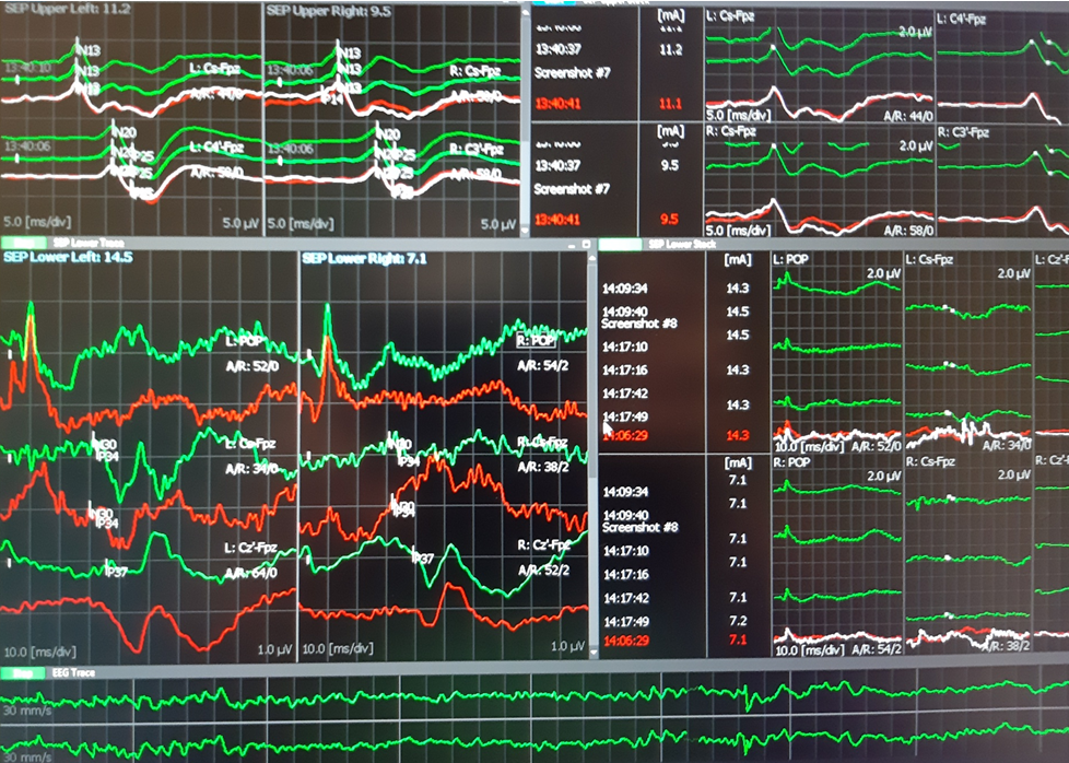
SSEP were performed from lower tibialis nerve and recorded to FpZ-Cz´ and from median nerves to Fpz-C3´ and Fpz-C4´. Both were controlled at popliteal fossa level (TN) and spinal C7 (TN and MN).
D wave was obtained rotral and caudal to the defect with D-wave electrodes after stimulation at 1Hz continuously (199V)
TOF was used from rioght median nerve to right APB.  was 100-99% during the entire surgery.
EEG was analized from C4´-Fpz and Fpz-C3´channels.

Results:
At the beginning of the surgery MEP was obtained in the upper limbs and left lower limb muscles. Right AH muscle was very difficult to elicit with 890 V, while TA in the right side was not obtained. At the end of the surgery the MEP were similar with those obtained at the beginning.

SSEP showed normal latencies during all the procedure. At the middle of the surgery, SSEP from right TN decreased 10-20% in amplitude. Rapidly we confirmed the decreased TA when asked the anaesthesiologist. After 5 minutes the SSEP recovered baseline values.

D-Wave was unchanged during all the spine manipulations and the defect corrective procedures.

Conclusion:
The IONM was successful and all modalities showed normal evolution. We do not expect sensory or motor new neurological deficits.

Technical data:
-Surgery duration 4,5 hours.
-Material: 11 subdermal needle paired electrodes, 4 monopolar subdermal needle electrodes, 2 D-wave electrodes, 8 Cork-screw electrodes, 1 ground electrode.

Neurophysiology Plus Iceland © 2019


Meningocele at T5 level.


 After surgery.
 SSEP upper and lower limbs

 MEP in lower limbs

 D wave: left rostral and right caudal
D wave: upper trace rostral, lower trace caudal. Latency 5-6 ms and amplitude 8uV.
The reference electrode was placed between two electrodes. (phase reversed peak).
It would be useful to reference both electrodes to a proximal sub-dermal well inserted electrode.

Monday, 26 February 2018

Cerebellopontine angle (CPA) mass IONM

by Neurophysiology Plus Iceland © 2018


IONM in 31 year-old male patient with 43/41mm right cerebello-pontine angle (CPA)  tumor
(e-Bulletin report)

We performed the following modalities:
Transcranial MEP, Corticogeniculate MEP (CoMEP), SSEP from both tibialis nerve, EEG, mapping cranial nerves (accesorius, hypoglossus, glosopharingeal, facialis ), BAEP, TOF, Blink Reflex.
Results: All modalities could be performed with exception of blink reflex which was not elicited.
Incidents: At the end of the surgery we observed decrement of right facialis (Orbicular oris muscle), CoMEP decrement of more than 80% which did not recover throughout the rest of the surgery. Surgeons explained that "there was a bleeding around the nerve". They started cooling,  irrigate and "treat" the lesion.  Mapping showed also partial decrement, but recovered after 20 minutes when stimulation was performed proximally.
Our protocol for corticobulbar MEP was double train: 1st train formed by 5 stimuli with 50 ms duration (ISI 2ms), 2nd train (single pulse 50 ms)  ITI (inter-train interval) 40 ms.
LIMIT: The assessment was possible for the right Orbicularis oris muscle and slightly for the right Orbicularis oculi muscle. No responses were obtained from the left muscles (we didn´t increase the stimulus intensity to look for the better Threshold-level ) and we consider the absence of other muscle MEP ipsilaterally (e.g. mentalis) as a serious limit of interpretation.


Right Orbicularis oris Corticogeniculate MEP decrement


Mapping with 0,3 mA baseline

Mapping with 0,86 mA (after the decrement was seen in CoMEP)

Conclusions:
Ø  All modalities with exception of  CoMEP showed similar findings at the end as at the beginning of the surgery. R1 (Trigeminofacial reflex) was not possible to elicit during this surgery.
Ø  Mapping was useful to drive the surgery moments before debulking and after the surgical removal of the tumor.  We expect partial facial nerve dysfunction (temporary deficit) in the right side.
Ø  24h after surgery, the patient showed 60% function of facial nerve preserved, Grade III (of VI) on House-Brackmann. Other VIII, IX and XII monitored cranial nerves didn´t show deficit 24 h after the surgery.
Ø  CoMEP as a measure of corticogeniculate motor tracts with different and variable assessment protocols should be considered as mandatory when trigeminofacial reflex (R1) cannot be monitored and the interpretation of the amplitude loss should be verified with anesthesist, neurosurgeons and with T-L technique (Calancie B, 2017).

Reference:
Intraoperative Neuromonitoring of CPA mass by Alba León Jorba & Ovidiu C. Banea (Oct 2014 IMGB)