Showing posts with label Neurophysiology Plus. Show all posts

Thursday, 24 January 2019

D wave IONM in Iceland. Third case: Meningocele


IONM for medular herniation at T5 level Use of D-wave 

HERE

Ovidiu C. Banea, Halldór Skúlasson, Ingvar H. Ólafsson, Aron D. Jónasson and Eysteinn Ívarsson

52 y.o. with meningocele.

Modalities:

MEP with direct cortical stimulation (560 V to the left and 890 V to the right) with train of five from C1-C2 and C3-C4 to:
- Right EDC, APB, TA, AH
- Left APB, TA, AH
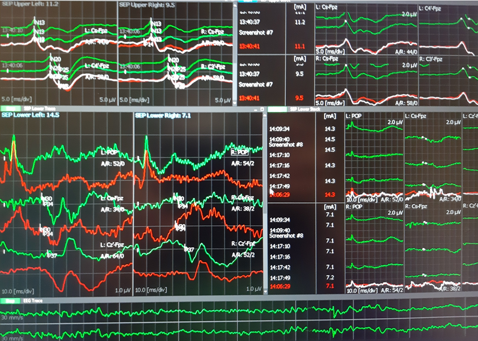
SSEP were performed from lower tibialis nerve and recorded to FpZ-Cz´ and from median nerves to Fpz-C3´ and Fpz-C4´. Both were controlled at popliteal fossa level (TN) and spinal C7 (TN and MN).
D wave was obtained rotral and caudal to the defect with D-wave electrodes after stimulation at 1Hz continuously (199V)
TOF was used from rioght median nerve to right APB.  was 100-99% during the entire surgery.
EEG was analized from C4´-Fpz and Fpz-C3´channels.

Results:
At the beginning of the surgery MEP was obtained in the upper limbs and left lower limb muscles. Right AH muscle was very difficult to elicit with 890 V, while TA in the right side was not obtained. At the end of the surgery the MEP were similar with those obtained at the beginning.

SSEP showed normal latencies during all the procedure. At the middle of the surgery, SSEP from right TN decreased 10-20% in amplitude. Rapidly we confirmed the decreased TA when asked the anaesthesiologist. After 5 minutes the SSEP recovered baseline values.

D-Wave was unchanged during all the spine manipulations and the defect corrective procedures.

Conclusion:
The IONM was successful and all modalities showed normal evolution. We do not expect sensory or motor new neurological deficits.

Technical data:
-Surgery duration 4,5 hours.
-Material: 11 subdermal needle paired electrodes, 4 monopolar subdermal needle electrodes, 2 D-wave electrodes, 8 Cork-screw electrodes, 1 ground electrode.

Neurophysiology Plus Iceland © 2019


Meningocele at T5 level.


 After surgery.
 SSEP upper and lower limbs

 MEP in lower limbs

 D wave: left rostral and right caudal
D wave: upper trace rostral, lower trace caudal. Latency 5-6 ms and amplitude 8uV.
The reference electrode was placed between two electrodes. (phase reversed peak).
It would be useful to reference both electrodes to a proximal sub-dermal well inserted electrode.

Monday, 20 November 2017

Neurophysiology Plus Iceland is represented at TMS-workshop in Denmark

by Ovidiu C. Banea


Questions for TMS research scientific community in Denmark

From November 22nd to November 24th 2017, Danish Research Center for Magnetic Resonance (DRCMR) will host a new TMS workshop with special focus on multimodal combinations of TMS with other neuroimaging techniques (EEG-TMS, fMRI-TMS). 
DRCMR is located in Hvidovre Hospital, a university hospital located at 9 km from Copenhagen which is administered by the Capital Region of Denmark.
Neurophysiology Plus will be represented at this meeting but also during the 20th to 22nd period for a better understanding of the center facilities, protocols used and technical equipment. 
We look mostly to have a valuable and critical analysis input from the team leaded by Prof Dr Hartwig Roman Siebner on the Icelandic proposed study. 
In this proposed clinical applied research project members of Neurophysiology Unit and Neurosurgery department from Reykjavik University and National University Hospital of Iceland are trying to analyze if TMS-EEG modality can be used or not to assess functional cortical tissue and brain effective connectivity in patients with brain tumors. 
In Iceland, another simple technique, TMS motor evoked potentials (TMS-EMG) started to be used for preoperative mapping in 2016. We set and marked the position for the intraoperative direct stimulation (IONM) as in the nineties when this technique was described. 
On the beginning of November 2017 neurosurgery department was interested on this procedure of preoperative mapping with neuronavigation. Again we used the available devices and we were able to map motor hotspots of the upper limb and speech area in a healthy subject. On 28th of November the team will investigate and perform motor and speech mapping in two patients with brain tumors located in eloquent areas of the brain. It will be for the first time that neuronavigated mapping is applied and used for the Icelandic brain tumor patients.

And the question remains: Is there a reason to believe that TEPs (TMS-EEG evoked potentials) can be used to assess better the "non-eloquent" brain cortical tissue and give a better map of the non-affected brain areas in order to avoid new post-intervention neurological deficit in patients with brain tumors ?