Showing posts with label Iceland. Show all posts

Friday, 13 December 2019

Road map for TMS and schizophrenia project in Iceland

Reykjavík University, Iceland

by Ovidiu C. Banea

On June 2018 a project dedicated to schizophrenia patients with auditory verbal hallucinations and their treatment with trans-cranial magnetic stimulation started in Iceland. 
The perfect coordination between researchers from Kleppur Psychiatric Hospital, Biomedical and Neural Engineering Institute at Reykjavík University, School of Business - Department of Psychology, National Institute of Neurological Disorders and Stroke (NINDS) Bethesda, United States and Clinical Neurophysiology Unit from National University Hospital - Landspítali Iceland made possible the recruitment and treatment of 12 patients with consequent analyses with multi-metric approach.
Measurements of the brain activity was performed before and after the treatment with P50, N100-P300 complex, resting state and auditory motor-task with 256 High Density-EEG recordings, cortical silent period and psychometric scales.
The team could organize and present their methodology, results and preliminary data in 14 original works during several local and international meetings in France, Japan, Poland, Portugal and Austria (see all works here).


On 13th of December we met at Reykjavik University and we discussed the detailed of the most important aspects of the AVH-TMS Icelandic Clinical trial - publishing the results in a peer review international journals. Two teams were formed, one at Reykjavik University and one at University National Hospital. The first team will process event related potential results at BNE and the second team will put together all data obtained at Fossvogur Landspítali and at Kleppur Psychiatric Hospital. By the end of January 2020 the process structure and software analyses will start.

Thursday, 24 January 2019

D wave IONM in Iceland. Third case: Meningocele


IONM for medular herniation at T5 level Use of D-wave 

HERE

Ovidiu C. Banea, Halldór Skúlasson, Ingvar H. Ólafsson, Aron D. Jónasson and Eysteinn Ívarsson

52 y.o. with meningocele.

Modalities:

MEP with direct cortical stimulation (560 V to the left and 890 V to the right) with train of five from C1-C2 and C3-C4 to:
- Right EDC, APB, TA, AH
- Left APB, TA, AH
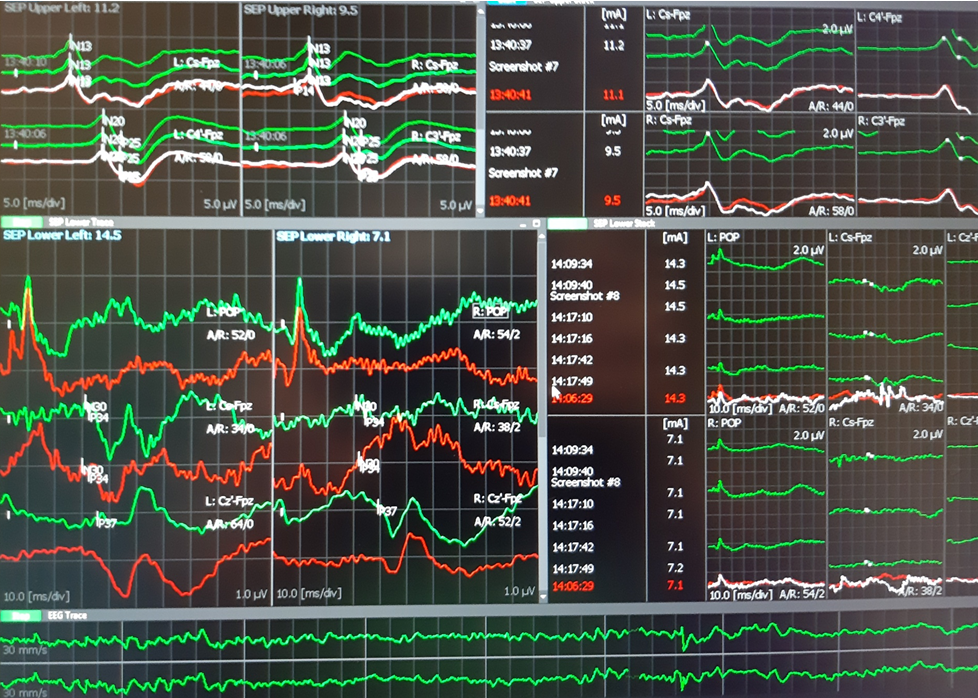
SSEP were performed from lower tibialis nerve and recorded to FpZ-Cz´ and from median nerves to Fpz-C3´ and Fpz-C4´. Both were controlled at popliteal fossa level (TN) and spinal C7 (TN and MN).
D wave was obtained rotral and caudal to the defect with D-wave electrodes after stimulation at 1Hz continuously (199V)
TOF was used from rioght median nerve to right APB.  was 100-99% during the entire surgery.
EEG was analized from C4´-Fpz and Fpz-C3´channels.

Results:
At the beginning of the surgery MEP was obtained in the upper limbs and left lower limb muscles. Right AH muscle was very difficult to elicit with 890 V, while TA in the right side was not obtained. At the end of the surgery the MEP were similar with those obtained at the beginning.

SSEP showed normal latencies during all the procedure. At the middle of the surgery, SSEP from right TN decreased 10-20% in amplitude. Rapidly we confirmed the decreased TA when asked the anaesthesiologist. After 5 minutes the SSEP recovered baseline values.

D-Wave was unchanged during all the spine manipulations and the defect corrective procedures.

Conclusion:
The IONM was successful and all modalities showed normal evolution. We do not expect sensory or motor new neurological deficits.

Technical data:
-Surgery duration 4,5 hours.
-Material: 11 subdermal needle paired electrodes, 4 monopolar subdermal needle electrodes, 2 D-wave electrodes, 8 Cork-screw electrodes, 1 ground electrode.

Neurophysiology Plus Iceland © 2019


Meningocele at T5 level.


 After surgery.
 SSEP upper and lower limbs

 MEP in lower limbs

 D wave: left rostral and right caudal
D wave: upper trace rostral, lower trace caudal. Latency 5-6 ms and amplitude 8uV.
The reference electrode was placed between two electrodes. (phase reversed peak).
It would be useful to reference both electrodes to a proximal sub-dermal well inserted electrode.

Monday, 13 August 2018

VAFO project Iceland, QEEG, P50, P300 recordings

On 11th of August 2018 VAFO project started officially with the first healthy subject data acquirement in the Icelandic Center for Neurophysiology, Department of Biomedical Engineering, Reykjavik University.


Valerio Gargiulo, Fabio Barollo, Eysteinn Ívarsson and Martin Freiler


Dr Sigurjón Stefánsson gave us important insights on methodology of P50 and P300. 
Continuous averaging is still a desired EEG system option to avoid artifacts during recordings.

Thursday, 9 August 2018

VAFO project Iceland: P50, P300, QEEG objective measurements for schizophrenia patients

by Neurophysiology Plus Iceland

Reykjavik, 9th of August 2018

A team of young researchers leaded by biomedical engineer Dr Paolo Gargiulo, from Reykjavík University, Department of Biomedical Engineering performed successfully the first data acquiring for a healthy subject recording P300, P50 and dense array 256-channel EEG.

P300 was first used in Iceland by Sigurjón B. Stefánsson and Anna L. Möller since the nineties.
Scientific data was published in 1995 and 2001 and 2005.
In July 2017 P300 returned to Iceland in Clinical Neurophysiology Unit of Landspitali (here).
P50 was acquired first time in patient with tinnitus on 20th of February 2018 in a paired-stimulus
or conditioning-testing paradigm, (S1, conditioning click; S2, testing click; 500 ms inter-click interval; 10,000 ms inter-pair interval). This was a trial of 30 paired stimulus with sensory gating with stimulus in the left and observed from Cz-A2 (right cortex) and with absent wave and response from the left Cz-A1 channel when stimulus was applied for the right ear.

Today the team formed by:

Viktor Díar Jónasson, Clinical Psychology master student at the Department of Psychology, Reykjavik University
Fabio Barrolo, PhD student at Reykjavik University Iceland / Aston University, Birmingham, UK, Biomedical Engineering
Valerio Gargiulo, Research assistant at the Icelandic Center for Neurophysiology, legal advisor and EEG technologist at Department of Biomedical Engineering, Reykjavík University
Eysteinn Ívarsson, Psychologist and technologist at Clinical Neurophysiology Unit, National University Hospital of Iceland
Ovidiu Banea, PhD student at Reykjavík University, Department of Biomedical Engineering and Clinical Neurophysiologist MD at National University Hospital of Iceland

performed both P300 and P50 acquisition with dense array 256 channel EEG system at the Icelandic Center for Neurophysiology, Department of Biomedical Engineering, Reykjavík University

Video on YouTube


artifact removal process, 256 channels EEG raw data

256 brain P50 map and selection of the 4 representatives lines

Valerio Gargiulo, Viktor Díar Jónasson, Eysteinn Ívarsson

Fabio Barollo


P 50 experimental


P300 experimental

256 channel dense array EEG, set-up


Friday, 18 May 2018

Yanny and Laurel explanation

Two days ago a BBC news announce that the computer generated word "Laurel" produced an internet debate if this is heard as "laurel" or "yanny".


The sound specialists run to explain how the humans are losing the higher frequencies listening ability with the age and this why we might listen the world with lower frequencies as is would be "laurel".
I was listening the playback and for two days since the Head of Neurology Department and one colleague told me the story and the new internet debate news, I always had listen the "yanny" word.
30 minutes ago I started to listen "laurel" word.
Yes, I know the word was created by computer and is an artificial voice the human cannot reproduce. The sounds are fake and the frequency differences are very difficult to assess by a computer voice generator. I remember the applications when you press a word text and suddenly you could listen it. This happened more than 6-7 years ago. There was no tonality, no stops or pause between words, no comma. You had to do everything.
But what happened to me? Why did I hear "laurel".
I did something before changes occurred. And I said to me, yes.
The gabaergic neurons from my cortex are weaker (the inhibitory mechanisms of my cortex were diminished by the substance intake)  and the sub-cortical areas and temporal lobe auditory cortex or let us say the auditory biological neural network is now a little bit more "awake"  and can hear the lower frequency. Or the higher frequencies are perceived with more glutamatergic (excitatory neurons) mechanisms? Is this known?
Ono et al 2017 found that responses to pure tones of both inhibitory and excitatory classes of neurons were similar in their thresholds, response latencies, rate-level functions, and frequency tuning, but GABAergic neurons may have higher spontaneous firing rates.
The auditory phantom perception biological neural network (BNN) 
involved in the subjective tinnitus perception

Conclusion:
The debate is easy: We are in front of unknown auditory perception mechanisms of a fake *artificial* sound. It seems that all is in our brain and that the gabaergic inhibitory mechanisms are involved. As much as you approach the freedom of the deeper brain structures (by forming gaps in the cortex "analytical" system) you will listen differently the word.

by Neurophysiology Plus Iceland, 18.05.2018


Monday, 26 February 2018

Cerebellopontine angle (CPA) mass IONM

by Neurophysiology Plus Iceland © 2018


IONM in 31 year-old male patient with 43/41mm right cerebello-pontine angle (CPA)  tumor
(e-Bulletin report)

We performed the following modalities:
Transcranial MEP, Corticogeniculate MEP (CoMEP), SSEP from both tibialis nerve, EEG, mapping cranial nerves (accesorius, hypoglossus, glosopharingeal, facialis ), BAEP, TOF, Blink Reflex.
Results: All modalities could be performed with exception of blink reflex which was not elicited.
Incidents: At the end of the surgery we observed decrement of right facialis (Orbicular oris muscle), CoMEP decrement of more than 80% which did not recover throughout the rest of the surgery. Surgeons explained that "there was a bleeding around the nerve". They started cooling,  irrigate and "treat" the lesion.  Mapping showed also partial decrement, but recovered after 20 minutes when stimulation was performed proximally.
Our protocol for corticobulbar MEP was double train: 1st train formed by 5 stimuli with 50 ms duration (ISI 2ms), 2nd train (single pulse 50 ms)  ITI (inter-train interval) 40 ms.
LIMIT: The assessment was possible for the right Orbicularis oris muscle and slightly for the right Orbicularis oculi muscle. No responses were obtained from the left muscles (we didn´t increase the stimulus intensity to look for the better Threshold-level ) and we consider the absence of other muscle MEP ipsilaterally (e.g. mentalis) as a serious limit of interpretation.


Right Orbicularis oris Corticogeniculate MEP decrement


Mapping with 0,3 mA baseline

Mapping with 0,86 mA (after the decrement was seen in CoMEP)

Conclusions:
Ø  All modalities with exception of  CoMEP showed similar findings at the end as at the beginning of the surgery. R1 (Trigeminofacial reflex) was not possible to elicit during this surgery.
Ø  Mapping was useful to drive the surgery moments before debulking and after the surgical removal of the tumor.  We expect partial facial nerve dysfunction (temporary deficit) in the right side.
Ø  24h after surgery, the patient showed 60% function of facial nerve preserved, Grade III (of VI) on House-Brackmann. Other VIII, IX and XII monitored cranial nerves didn´t show deficit 24 h after the surgery.
Ø  CoMEP as a measure of corticogeniculate motor tracts with different and variable assessment protocols should be considered as mandatory when trigeminofacial reflex (R1) cannot be monitored and the interpretation of the amplitude loss should be verified with anesthesist, neurosurgeons and with T-L technique (Calancie B, 2017).

Reference:
Intraoperative Neuromonitoring of CPA mass by Alba León Jorba & Ovidiu C. Banea (Oct 2014 IMGB)

Monday, 20 November 2017

Neurophysiology Plus Iceland is represented at TMS-workshop in Denmark

by Ovidiu C. Banea


Questions for TMS research scientific community in Denmark

From November 22nd to November 24th 2017, Danish Research Center for Magnetic Resonance (DRCMR) will host a new TMS workshop with special focus on multimodal combinations of TMS with other neuroimaging techniques (EEG-TMS, fMRI-TMS). 
DRCMR is located in Hvidovre Hospital, a university hospital located at 9 km from Copenhagen which is administered by the Capital Region of Denmark.
Neurophysiology Plus will be represented at this meeting but also during the 20th to 22nd period for a better understanding of the center facilities, protocols used and technical equipment. 
We look mostly to have a valuable and critical analysis input from the team leaded by Prof Dr Hartwig Roman Siebner on the Icelandic proposed study. 
In this proposed clinical applied research project members of Neurophysiology Unit and Neurosurgery department from Reykjavik University and National University Hospital of Iceland are trying to analyze if TMS-EEG modality can be used or not to assess functional cortical tissue and brain effective connectivity in patients with brain tumors. 
In Iceland, another simple technique, TMS motor evoked potentials (TMS-EMG) started to be used for preoperative mapping in 2016. We set and marked the position for the intraoperative direct stimulation (IONM) as in the nineties when this technique was described. 
On the beginning of November 2017 neurosurgery department was interested on this procedure of preoperative mapping with neuronavigation. Again we used the available devices and we were able to map motor hotspots of the upper limb and speech area in a healthy subject. On 28th of November the team will investigate and perform motor and speech mapping in two patients with brain tumors located in eloquent areas of the brain. It will be for the first time that neuronavigated mapping is applied and used for the Icelandic brain tumor patients.

And the question remains: Is there a reason to believe that TEPs (TMS-EEG evoked potentials) can be used to assess better the "non-eloquent" brain cortical tissue and give a better map of the non-affected brain areas in order to avoid new post-intervention neurological deficit in patients with brain tumors ?

Thursday, 2 November 2017

Neuronavigated TMS in Iceland

2nd of November 2017


After 11 months since the first awake craniotomy was performed in Iceland, medical and technical staff from Neurosurgery Department and Clinical Neurophysiology Unit of the Icelandic National  University Hospital #Landspitali started new testing protocol for the patients with lesions located in eloquent areas of the brain.  
The neuronavigated transcranial magnetic stimulation (neuronavigated-TMS) was initiated with a pilot study of a healthy subject on 1st of November 2017 to find motor "hot-spots" and "speech area" (by inducing the speech arrest) in total relaxation and safety.  
When the team will perform the test in patients, the functional areas will be saved with Talairach space, a 3-dimensional coordinate system of the brain structures, into the same neuronavigation system used by neurosurgeons in the operating room with the aim to avoid their damage during the surgical procedures.
On 13th of July 2017 the team tried this set-up on a phantom head in the operating room theater (here). 
Many thanks to Dr Ingvar Hákon Ólafsson (Neurosurgery Department) for his interest and collaboration, to Aron Dalin Jónasson (TMS Clinical Neurophysiology Unit technician) to support the MRI procedure and the pilot noninvasive TMS neuronavigation, Ágúst H. Guðmundsson (Intermedica & Medtronic) and to all Neurophysiology Unit medical and technical staff.


Friday, 29 September 2017

TMS-EEG evoked potentials (TEPs) in Iceland

29th of Sept 2017 Reykjavik Iceland

TMS-EEG evoked potentials (TEP) were performed during a joint meeting of Clinical Neurophysiology Unit team (Neurophysiology Plus Iceland) and Icelandic Center of Clinical Neurophysiology from Reykjavik University.
The meeting was organized by Assist Prof Dr Paolo Gargiulo (Director of Institute of Biomedical and Neural Engineering, Reykjavik University & Landspitali) and had as participants Dr Magnús Kjartan Gíslason & MSc Thorsteinn Geirsson (NeckCare), Aron Dalin Jónasson MSc, Hildigunnur Katrinardóttir MSc & Ovidiu C. Banea MD (Neurophysiology Lab Landspitali and Reykjavik University) and Egill Axfjörður Friðgeirsson, PhD Student University of Amsterdam.


First TMS-EMG was performed to achieve the correct out of maximum TMS intensity necessary to evoke MEP into hand thenar muscles


Using 100% RMT TMS-EEG evoked potentials were recorded. This trial was performed on experimental basis within the expert team in a healthy subject who previously accepted the test. Another 15-20 voluntary healthy subjects will be tested on both TMS-EMG and TMS-EEG protocols in accordance with World Medical Association (WMA) Declaration of Helsinki, a statement of ethical principles for medical research involving human subjects.

The future joint applied science clinical study will be developed after two international specific trainings and meetings in Denmark (Nov 2017) and France (January 2018). The aim is to assess biological neural networks (BNN) in patients with brain tumors and symptomatic epilepsy as a preoperative safety assessment of the functional brain tissue and effective connectivity in order to improve actual procedures (motor  & speech mapping and fMRI) and avoid new neurological deficit. Main collaborators of this challenging Icelandic medical research and clinical study are Neurosurgery Department of National University Hospital of Iceland (Drs Ingvar Hakon Ólafsson & Elfar Ulfarsson) and Neurosurgery Department of del Mar Hospital Barcelona, Spain (Dr Gerard Conesa). Scientific support and research specific feedback is given by Dr Eric Wassermann (NINDS/NIH; Bethesda, United States) and Prof Dr Elías Ólafsson (Head of Neurology Department, National University Hospital of Iceland).

Wednesday, 26 July 2017

Mixed silent period (MSP) and cutaneous silent period(CSP) in diagnostic of a severe carpal tunnel syndrome

Reykjavik 26th of July 2017

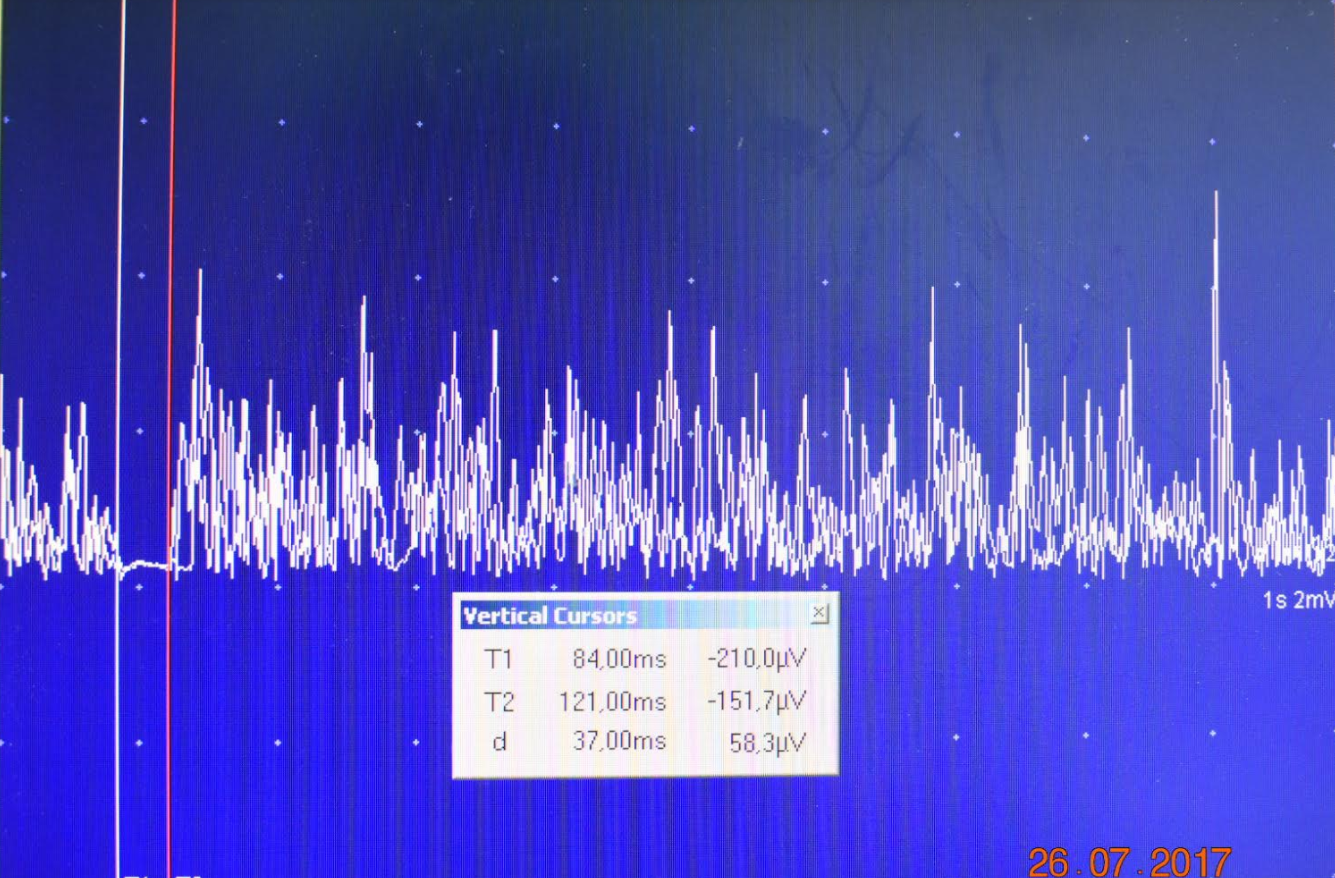
Cutaneous silent period can be preserved in entrapment neuropathies (KofIer et al 2003). It is known that the more severe the neuropathy is, the more impairment of A-delta fibers can be found (Duarte et al 2016). In patient with severe CTS the mean onset latency was increased to 85.0 ms (SD 8.7 ms, P < 0.01) Silvpauskaite et al 2005.

CASE REPORT (herePatient 66 year-old with motor weakness in hands and hypoesthesia

This case was published in Neurophysiology Plus Iceland informal group e-Bulletin (here)

Right median nerve mixed and cutaneous (palm) silent period

Cutaneous Silent Period from 2 digit sensory branches (ring electrodes)

Cutaneous Silent Period from 2 digit sensory branches in examinator (OCB), normal values

After we diagnosed very severe CTS in the right side and severe CTS in the left we performed A-delta & alpha motorneurons driven CSP from 2 digit and there was no clear inhibition of the voluntary contraction (onset 107ms). Then, we checked again from the palm-thenar region and we observed CSP with onset 100ms, end 155 ms and 55ms duration. Also a clear silent period was obtained after stimulation from the wrist, the mixed median nerve and record from abductor pollicis brevis.
Conclusion: Sensory fibers (small and fast) are the last in be damaged and the injury do not occurred at the wrist carpal tunnel as it happened with axonal loss of the large sensory fibers.  At this moment there is not clear if those fibers are only the A-delta or there might be another sensory input by direct electrical stimulation of other fiber type (proprioceptors?, muscle spindles?)


Nerve Conduction Studies: 2017 © Neurophysiology Plus Iceland


Monday, 24 July 2017

P300 returns to Iceland

Reykjavik 24.07.2017

by Neurophysiology Plus

The P300 (P3) wave is an event related potential (ERP) component elicited in the process of decision making.

Since the mid-1980s, one of the most discussed uses of ERPs such as the P300 is related to lie detection. In a proposed "guilty knowledge test" (Farwell & Smith, 2001) a subject is interrogated via the oddball paradigm much as they would be in a typical lie-detector situation. This practice has recently enjoyed increased legal permissibility while conventional polygraphy has seen its use diminish, in part owing to the unconscious and uncontrollable aspects of the P300.


Photo: Aron Dalin Jónasson