Wednesday 26 July 2017

Mixed silent period (MSP) and cutaneous silent period(CSP) in diagnostic of a severe carpal tunnel syndrome

Reykjavik 26th of July 2017

Cutaneous silent period can be preserved in entrapment neuropathies (KofIer et al 2003). It is known that the more severe the neuropathy is, the more impairment of A-delta fibers can be found (Duarte et al 2016). In patient with severe CTS the mean onset latency was increased to 85.0 ms (SD 8.7 ms, P < 0.01) Silvpauskaite et al 2005.

CASE REPORT (herePatient 66 year-old with motor weakness in hands and hypoesthesia

This case was published in Neurophysiology Plus Iceland informal group e-Bulletin (here)

Right median nerve mixed and cutaneous (palm) silent period

Cutaneous Silent Period from 2 digit sensory branches (ring electrodes)

Cutaneous Silent Period from 2 digit sensory branches in examinator (OCB), normal values

After we diagnosed very severe CTS in the right side and severe CTS in the left we performed A-delta & alpha motorneurons driven CSP from 2 digit and there was no clear inhibition of the 
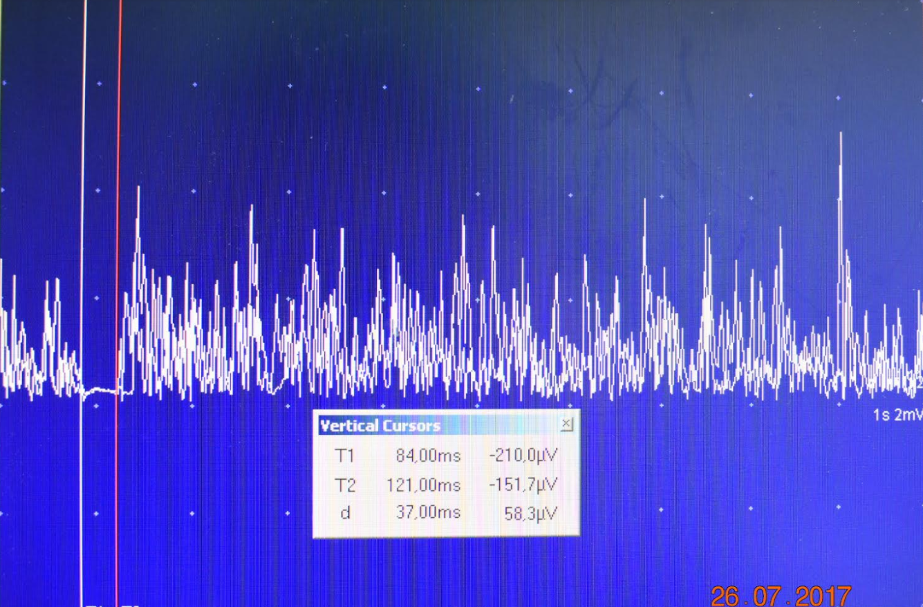
voluntary contraction (onset 107ms). Then, we checked again from the palm-thenar region and we observed CSP with onset 100ms, end 155 ms and 55ms duration. Also a clear silent period was obtained after stimulation from the wrist, the mixed median nerve and record from abductor pollicis brevis.
Conclusion: Sensory fibers (small and fast) are the last in be damaged and the injury do not occurred at the wrist carpal tunnel as it happened with axonal loss of the large sensory fibers.  At this moment there is not clear if those fibers are only the A-delta or there might be another sensory input by direct electrical stimulation of other fiber type (proprioceptors?, muscle spindles?)


Nerve Conduction Studies: 2017 © Neurophysiology Plus Iceland


Monday 24 July 2017

P300 returns to Iceland

Reykjavik 24.07.2017

by Neurophysiology Plus

The P300 (P3) wave is an event related potential (ERP) component elicited in the process of decision making.

Since the mid-1980s, one of the most discussed uses of ERPs such as the P300 is related to lie detection. In a proposed "guilty knowledge test" (Farwell & Smith, 2001) a subject is interrogated via the oddball paradigm much as they would be in a typical lie-detector situation. This practice has recently enjoyed increased legal permissibility while conventional polygraphy has seen its use diminish, in part owing to the unconscious and uncontrollable aspects of the P300.


Photo: Aron Dalin Jónasson

Thursday 20 July 2017

Tinnitus experimental treatment with repetitive TMS in Iceland

20 of July 2017, Reykjavik

by Neurophysiology Plus

Experimental treatment with repetitive TMS (rTMS) for subjective tinnitus is a challenging option. Preliminary research data on rTMS has yielded mixed results, especially when rTMS patients are compared to patients in a placebo test group (American Tinnitus Association). 
Low frequency ( LF 1 Hz) rTMS unilaterally applied to temporal or temporoparietal cortical areas can interact with an abnormal hyperactivity of auditory cortices that may constitute the neural correlate of tinnitus perception. Literature data showed that this type of rTMS protocol has a possible therapeutic efficacy (Level C recommendation) in this clinical condition (J.-P. Lefaucheur et al.2014 / Clinical Neurophysiology 125, page 2177).
In Iceland, our first patient started the 10 sessions treatment with 1200 daily pulses 100%RMT applied in 60 series of 20 pulses (1Hz frequency) on 17th of July 2017. Location of coil was set to stimulate between T3 and C3/T5 using 10-20 international EEG system (Lee H.Y. et al 2013). 
We use the same protocol used in Department of Otorhinolaryngology - Head and Neck Surgery, Kyung Hee University School of Medicine, Seoul (Lee H.Y. et al 2013Clinical and Experimental Otorhinolaryngology Vol. 6, No. 2: 63-67, June 2013). To assess the progression of a possible rTMS effect on tinnitus perception we used Tinnitus Handicap Inventory THI questionnaire and Visual Analogue Scale for both tinnitus annoyance and loudness (here). 

Primary Auditory Cortex: In humans, the primary auditory cortex is located in the transverse temporal gyri (of Heschl) of the medial aspect of the superior temporal gyrus. Brodmann’s area 42 is the auditory association area. Together, Brodmann’s areas 41 and 42 are called the A-1 region and receive projections from the medial geniculate nucleus (geniculotemporal fibers or auditory radiations). The tonotopic organization in the auditory relay nuclei is maintained in the auditory cortex as well. One of the secondary auditory areas includes Wernicke’s area, which is important for the interpretation of the spoken word.


20.07.2017 Clinical Neurophysiology Unit, Reykjavik
Photo: Aron Dalin Jónasson

Tuesday 18 July 2017

Preoperative Neuronavigation with TMS using operating room system in Iceland

Reykjavik, 13th of July 2017

by Neurophysiology Plus

To avoid a new neurological deficit after a brain growing tumor surgery located in or near eloquent brain areas (speech, motor, visual cortex) the surgeon uses now intraoperative neuromonitoring complementary procedures performed together with clinical neurophysiology team in the operating room.

In 1998, Sawaya et al published the tumor functional grade classification after a scheme developed at the Anderson Cancer Center in Houston Texas based on the tumor location relative to the brain function as following: Grade I non-eloquent brain: Frontal or temporal polar regions, Right parieto-occipital regions, Cerebellar hemispheres lesions; Grade II near eloquent brain: Near motor or sensory cortex, Near calcarine fissure, Near speech center, Corpus callosum, Near dentate nucleus, Near brainstem, including supplementary motor area if was investigated with preoperative magnetic resonance; Grade III eloquent brain: Motor/Sensory cortex, Visual center, Speech center, Internal capsule, Basal ganglia, Hypothalamus/thalamus, Brainstem, Dentate nucleus.

The preoperative studies used to identify the structure and the "function" of the eloquent brain are MRI, fMRI, CT, DatScan, SPECT, PET, TMS. From all these TMS-EMG and TMS-EEG modalities are used for motor mapping and speech mapping or EEG temporal related evoked potentials. The results, the identified motor hot-spots or speech area are then saved and used in operating room by neurosurgeon.

Source: Medtronic

Neuro-Navigated TMS preoperative mapping using same operating room system
13th of July 2017

If eloquent brain areas and motor cortex hot-spots with Talairach coordinates are already set on the patient MRI (structural model of the brain) before surgery the neurophysiology and neurosurgery team will use them when proposing the surgery and during the resection of tumors or abnormal (non-functional) tissues.


In Iceland, preoperative motor mapping started in January 2017 (here) and recently a team formed by Ingvar Hakón Ólafsson, Aron Björnsson, Ágúst H. Guðmundsson (Intermedica/Medtronic), David B., Aron Dalin Jónasson and Ovidiu C. Banea could prove the combination of operating room neuronavigation system (Medtronic) with TMS motor mapping and Intraoperative device (Nim Eclipse Medtronic).
The pilot study was successful and was performed in surgery room. In close future the preoperative neuro-navigated mapping will be used for the proposed patients before surgery time.