
HERE
Ovidiu C. Banea, Halldór Skúlasson, Ingvar H. Ólafsson, Aron D. Jónasson and Eysteinn Ívarsson
52 y.o. with meningocele.
Modalities:
MEP with direct cortical stimulation (560 V to the left and 890 V to the right) with train of five from C1-C2 and C3-C4 to:
- Right EDC, APB, TA, AH
- Left APB, TA, AH
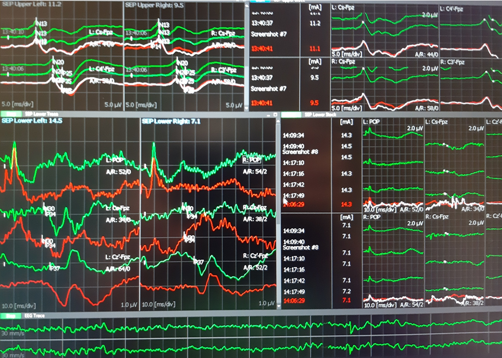
SSEP were performed from lower tibialis nerve and recorded to FpZ-Cz´ and from median nerves to Fpz-C3´ and Fpz-C4´. Both were controlled at popliteal fossa level (TN) and spinal C7 (TN and MN).
D wave was obtained rotral and caudal to the defect with D-wave electrodes after stimulation at 1Hz continuously (199V)
TOF was used from rioght median nerve to right APB. was 100-99% during the entire surgery.
EEG was analized from C4´-Fpz and Fpz-C3´channels.
Results:
At the beginning of the surgery MEP was obtained in the upper limbs and left lower limb muscles. Right AH muscle was very difficult to elicit with 890 V, while TA in the right side was not obtained. At the end of the surgery the MEP were similar with those obtained at the beginning.
SSEP showed normal latencies during all the procedure. At the middle of the surgery, SSEP from right TN decreased 10-20% in amplitude. Rapidly we confirmed the decreased TA when asked the anaesthesiologist. After 5 minutes the SSEP recovered baseline values.
D-Wave was unchanged during all the spine manipulations and the defect corrective procedures.
Conclusion:
The IONM was successful and all modalities showed normal evolution. We do not expect sensory or motor new neurological deficits.
Technical data:
-Surgery duration 4,5 hours.
-Material: 11 subdermal needle paired electrodes, 4 monopolar subdermal needle electrodes, 2 D-wave electrodes, 8 Cork-screw electrodes, 1 ground electrode.
Neurophysiology Plus Iceland © 2019

Meningocele at T5 level.


D wave: upper trace rostral, lower trace caudal. Latency 5-6 ms and amplitude 8uV.